MIT's xvr Turns X-Rays Into Real-Time Surgical Maps for Orthopedics and Neurosurgery
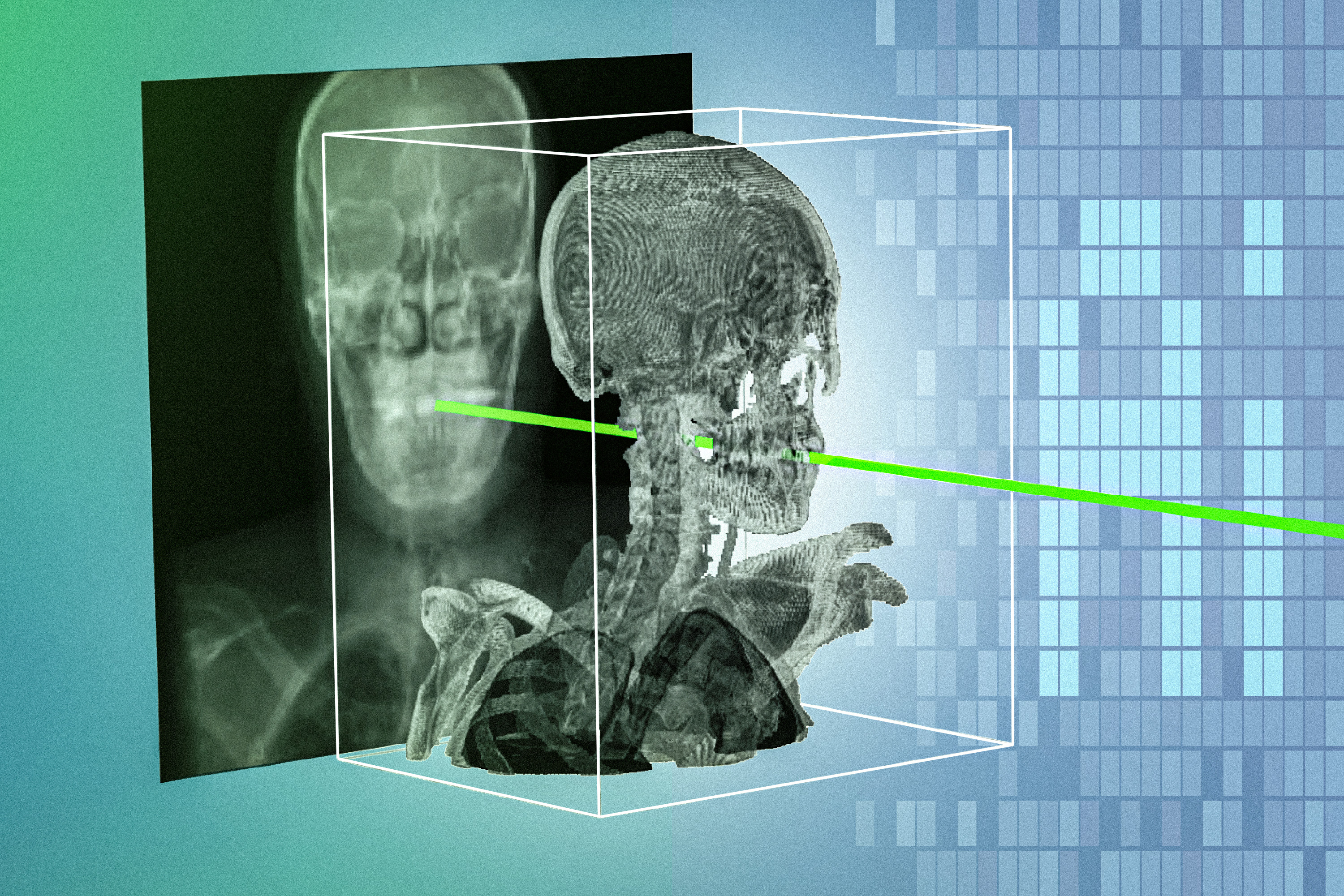
A patient-specific imaging method from MIT aims to sharpen X-ray guidance during minimally invasive spine, brain and bone procedures.
Researchers at MIT have introduced a new imaging method called xvr that uses a patient's own X-rays to build surgical navigation guidance, a step aimed at making minimally invasive operations safer and more precise. The technique is designed for fields where surgeons work through small openings and rely on fluoroscopy to see instruments inside the body, including orthopedics and neurosurgery. MIT News reported the development this week.
Minimally invasive surgery trades a large incision for a narrow corridor, which spares tissue but strips the surgeon of direct sight. X-ray fluoroscopy fills that gap in the operating room, yet it delivers flat, two-dimensional projections of three-dimensional anatomy, and it exposes both patient and staff to repeated radiation doses over the course of a procedure. Errors in placing screws, implants or catheters carry real consequences in the spine and skull, where critical nerves and vessels sit millimeters away from the target.
What separates xvr from conventional navigation is the word patient-specific. Rather than fitting every case to a generic atlas or requiring a preoperative CT scan, the method derives its model from the individual's X-ray data, according to the MIT report. That matters for hospitals that already have X-ray equipment in place but lack the capital or workflow capacity for CT-based or MRI-based navigation suites, which are expensive and add setup time before the first incision.
The approach lands in a crowded field of image-guided surgery tools, where established systems from major device makers and a wave of surgical robotics startups compete for operating room space. Most of those platforms anchor themselves to preoperative scans and then register the patient to that data in the OR, a step that can drift as tissue shifts during the case. A method that reconstructs anatomy from intraoperative X-rays could reduce that mismatch if it holds up under clinical conditions.
The obvious next question is validation. MIT's announcement describes a technique, not a completed clinical trial, so the numbers that will decide its fate are still to come: accuracy against ground-truth anatomy, radiation dose compared with standard fluoroscopy, and performance across real patients rather than phantoms or retrospective datasets. Watch for peer-reviewed results and for whether any orthopedic or neurosurgery group signs on to test xvr in live cases.
If those trials show the method matches or beats existing navigation on accuracy while cutting radiation exposure, xvr could give community hospitals a cheaper route into image-guided spine and brain surgery. Until then, it remains a promising research result in a specialty where the bar for adoption is set by operating room workflow, surgeon trust and regulatory clearance, not by the elegance of the algorithm.